Loma Linda Dermatopathology
Dermatopathology/Dermatology Histopathology Library Reference
Disclaimer: The information on this website is provided for informational
and educational purposes by Fred F. Soeprono, M.D., Board-Certified
Dermatopathologist and Dermatologist. No warranty or
guarantee is provided as to the accuracy of the information provided on this website.
Consult a licensed medical doctor with any medical questions.
intravascular papillary endothelial hyperplasia
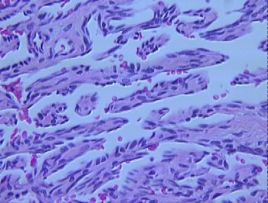
Histologic Features
- A fibrous pseudocapsule containing residual wall of preexisting vessels
- Papillary fronds lined by plump endothelial cells without pleomorphism or mitotic figures
- Collagenized cores developing from organized thrombi
- Fibrin and thrombus material with hemorrhage
- Spindle cells and scattered small vessels lined by plump endothelial cells (later stages)
Looking for more information,
high-resolution images,
or differential diagnoses on this entity?
Copyright 2012, Fred F. Soeprono, M.D. You are granted a limited, non-exclusive, revocable license to view the content on this website.
Copying, reproducing, or publishing the content on this website is strictly prohibited and not part of such license.

Dr. Soeprono's textbook is available on Amazon.com and includes detailed information on over 600 entities and includes four DVD diskettes with high-resolution images that provide a virtual dermatopathology reference and guide. 
|

Dr. Soeprono teaches and practices dermatopathology at Loma Linda University, School of Medicine, Department of Dermatology. He also practices dermatology at the Advanced Dermatology & Laser Center of Redlands, CA and dermatopathology privately. |
Alphabetized Index
View information about other entities available on this website.
This dermatopathology histopathology reference contains information about over 600 dermatopathology entities.
to over 600 entities in this dermatopathology/ histopathology library. |