Loma Linda Dermatopathology
Dermatopathology/Dermatology Histopathology Library Reference
Disclaimer: The information on this website is provided for informational
and educational purposes by Fred F. Soeprono, M.D., Board-Certified
Dermatopathologist and Dermatologist. No warranty or
guarantee is provided as to the accuracy of the information provided on this website.
Consult a licensed medical doctor with any medical questions.
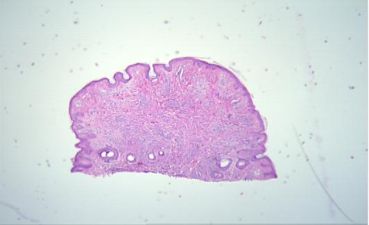
dysplastic nevus (stepsections)

Histologic Features
- Multiple sagittal stepsections were obtained through the specimen and mounted as diagrammed. Examination of the sections show an asymmetrical proliferation of slightly atypical melanocytes as nests and single cells at the DE junction, concentric fibroplasia of the papillary dermis, pigment incontinence and a lymphocytic infiltrate at the base. The surgical margins are ample and free of involvement
Looking for more information,
high-resolution images,
or differential diagnoses on this entity?
Copyright 2012, Fred F. Soeprono, M.D. You are granted a limited, non-exclusive, revocable license to view the content on this website.
Copying, reproducing, or publishing the content on this website is strictly prohibited and not part of such license.

Dr. Soeprono's textbook is available on Amazon.com and includes detailed information on over 600 entities and includes four DVD diskettes with high-resolution images that provide a virtual dermatopathology reference and guide. 
|

Dr. Soeprono teaches and practices dermatopathology at Loma Linda University, School of Medicine, Department of Dermatology. He also practices dermatology at the Advanced Dermatology & Laser Center of Redlands, CA and dermatopathology privately. |
Alphabetized Index
View information about other entities available on this website.
This dermatopathology histopathology reference contains information about over 600 dermatopathology entities.
to over 600 entities in this dermatopathology/ histopathology library. |